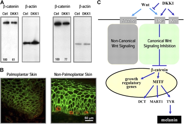
Figure 6.
Regulation of cell signaling intermediates by DKK1. (A) Expression of β-catenin was analyzed by Western blot in cell extracts obtained from melanocytes cocultured for 5 d with control- or DKK1-transfected fibroblasts (left) or from melanocytes treated for 3 h with or without 50 ng/ml DKK1 (right). β-actin is shown as a loading control. The numbers below the bands represent their quantitation as a percentage of control, corrected against the β-actin loading control. This experiment was performed four times with melanocytes and fibroblasts derived from different individuals with similar results. (B) Immunohistochemical studies were performed using biopsy specimens of palmoplantar and nonpalmoplantar skin. The expression of β-catenin was examined (stained green), and melanocytes were detected by localization of MART1 (stained red). (C) Scheme illustrating the potential mechanism by which DKK1 decreases melanocyte growth and differentiation.

