Figure 8.

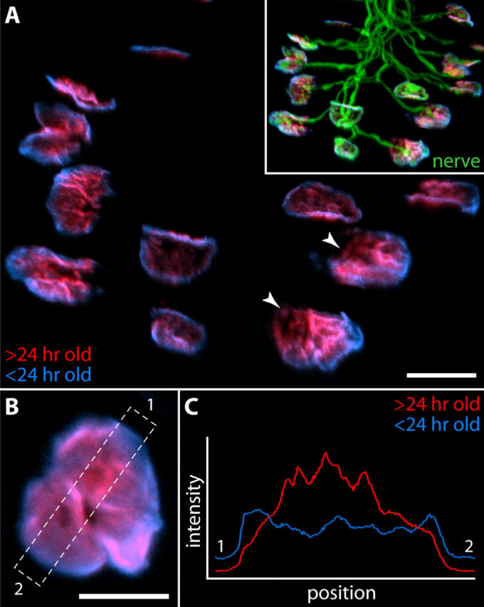
Circumferential growth of AChR clusters at the NMJ. (A) Btx was injected above the sternomastoid muscle of a postnatal day five pup that expressed YFP in its motor axons. After 1 d, the pup was killed, and unlabeled AChRs were stained with a second, spectrally distinct Btx conjugate. AChRs carrying the first tag (red) are found preferentially near the center of aggregates, whereas younger AChRs (blue) are concentrated at the periphery. Note that in “open” aggregates (arrowheads) new AChRs are preferentially concentrated along the convex margin. The nerve invariably enters the synapse from the opposite side (inset, overlay with YFP to show pattern of innervation). (B) High resolution image of a single junction labeled as above. (C) Relative intensities of the two labels in the region boxed in B. Bars: (A) 20 μm; (B) 10 μm.
