
Figure 9.
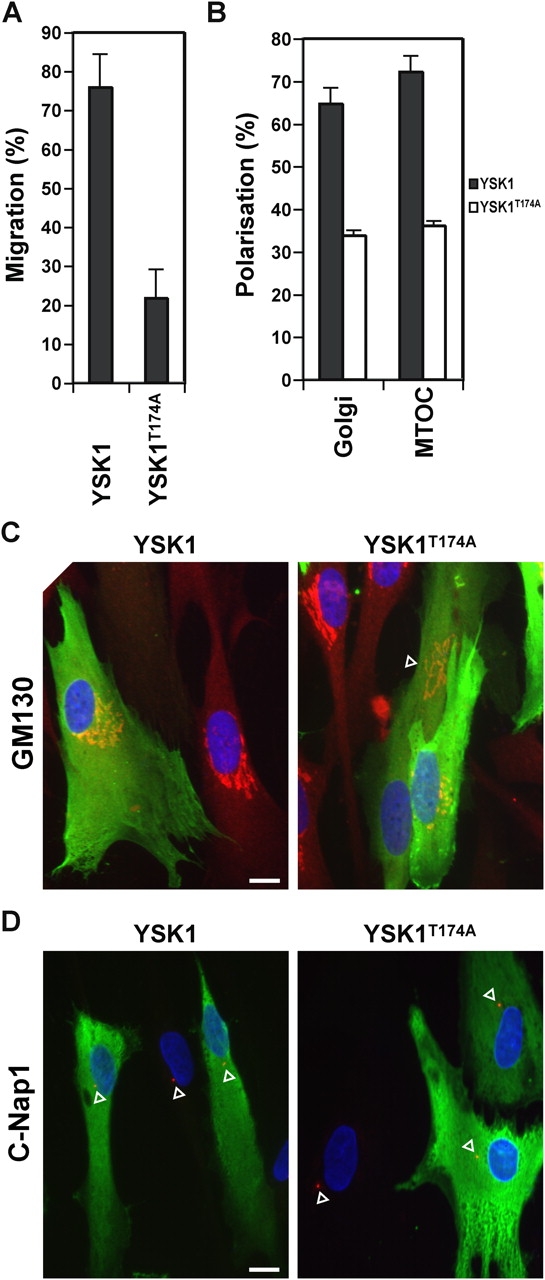
Golgi apparatus and centrosome polarization is inhibited by YSK1T174A. (A and B) Cells were microinjected with plasmids encoding myc-tagged YSK1 and YSK1T174A, fixed after 16 h, and stained with antibodies to GM130 or c-Nap1, and 9E10 monoclonal to the myc epitope. Bar graphs show the percentage of YSK1 or YSK1T174A expressing cells migrating at the wound edge or into the wound (n = 12), and the percentage of cells with Golgi apparatus and centrosomes (MTOC) polarized toward the wound edge (n = 4). (C and D) Images of cells expressing YSK1 or YSK1T174A (green) and stained for GM130 or c-Nap1 (red) are shown. The position of the wound corresponds to the bottom of the figure. Arrowheads indicate the position of the Golgi apparatus and centrosome. Bars, 10 μm.