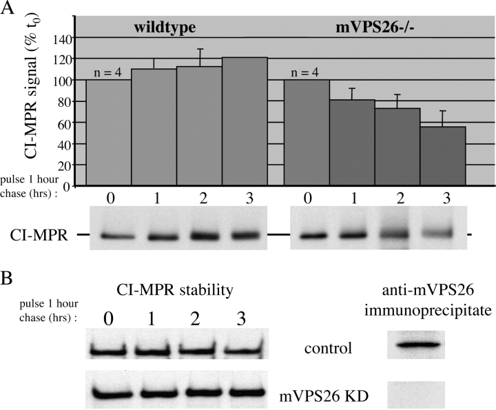
Figure 5.
The CI-MPR is unstable in the mVPS26 −/− cells. (A) Cells grown in 3-cm dishes were pulse labeled with [35S]methionine for 1 h and then chased for 0, 1, 2, or 3 h. The cells were lysed and the CI-MPR was recovered by immunoprecipitation and then subjected to SDS-PAGE and fluorography. The signal on the resulting film was quantified and expressed as a percentage of the signal at time 0. The data from four experiments were averaged together and are shown in the graph. The error bars are SDs. The bottom panels are representative of the data obtained and show the instability of the CI-MPR in the mVPS26−/− cells. (B) The CI-MPR stability experiment was repeated as described above using control and mVPS26 knock-down cells. There is no apparent instability of the CI-MPR after mVPS26 knock down.

