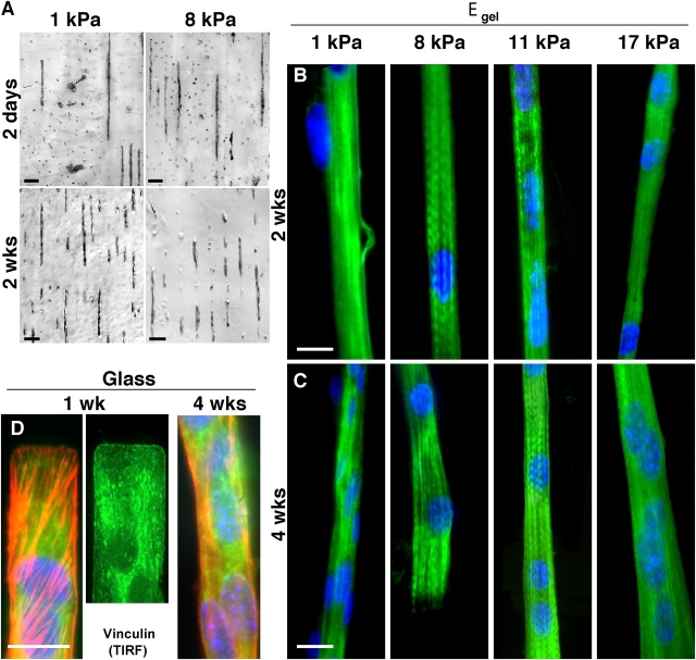
Figure 4.
Myotube striation is substrate stiffness dependent, whereas myocyte patterning is not. (A) By 2 d in culture, myoblasts align on the collagen patterns and begin to fuse into nascent myotubes. This occurs regardless of substrate elastic modulus and is sustainable in culture for weeks. Unbranched, isolated myotubes are consistently >90% of cells throughout the lifetime of all micropatterned cultures. Mouse primary cells (C57 derived) exhibit a similar behavior. (B and C) Several weeks after plating myoblasts on collagen-coated PA gels of varied stiffness or rigid glass, cells were stained for myosin (green), actin (not depicted), and the nucleus (blue) in order to assess cytoskeletal expression and organization. All substrates supported fusion into multi-nucleated myotubes, but after 2 or 4 wk only myotubes on gels of intermediate stiffness showed significant myosin striation. (D) On rigid glass, nascent myotubes showed robust arrays of stress fibers (F-actin is red) and tight substrate adhesion from vinculin by 1 wk and, as extrapolated from the PA gel results, myotubes showed no tendency to striate, even by 4 wk. Note the precision of the end-patterning on glass (see Materials and methods). Bars: (A) 100 μm; (B–D) 20 μm.

