Figure 6.
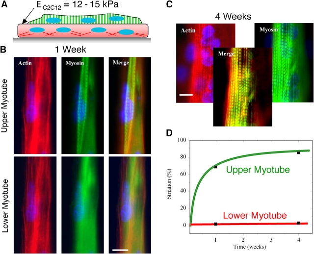
Myotube underlayers provide optimal stiffness for myotube striation. Myoblasts were plated in two sequential layers on patterned glass. (A) An initial, bottom layer of cells was added and is schematically represented as nascent myotubes with square ends “pinned” by stress fibers. Subsequent addition of cells generates an top myotube that progressively differentiates. Within 1 wk after plating (B) and even more prominent at 4 wk (C), the top layer of cells striate both F-actin and myosin although clear striation of F-actin seems slower. Merged images of the top myotube at 4 wk demonstrate actin-myosin colocalization. Bars, 10 μm. (D) Striated myotubes in the top layer, after 1 and 4 wk, respectively constituted 68% (n = 34) and 85% of the total myotube population (repeated in duplicate). Immunostaining of vinculin in cells on top of cells shows diffuse labeling and no profound focal adhesions (not depicted).

