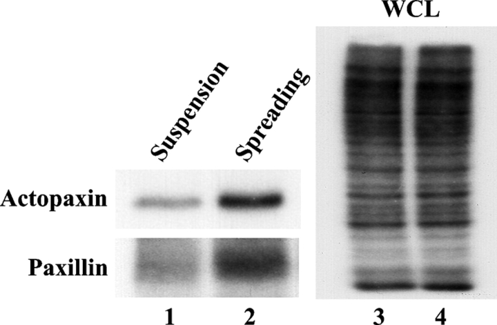
Figure 2.
Actopaxin is phosphorylated in vivo after cell adhesion. Cells were labeled with inorganic 32P and either held in suspension (lanes 1 and 3) or spread on collagen I–coated dishes for 2 h (lanes 2 and 4). Actopaxin (top) and paxillin (bottom) proteins were immunoprecipitated, resolved on SDS-PAGE, and visualized by autoradiography. WCL, whole cell lysate.

