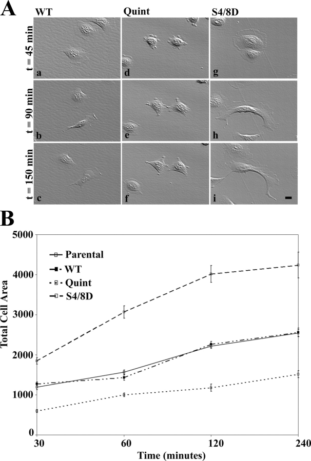
Figure 5.
Actopaxin phosphorylation mutants affect cell spreading. Actopaxin WT, Quint, and S4/8D cells were plated on collagen type 1–coated dishes and evaluated by time-lapse video microscopy. (A) Representative Hoffman illumination images were acquired at 45, 90, and 150 min after plating. Quint cells are poorly spread and lack lamellipodia at all time points (d-f) compared with WT (a–c), whereas the S4/8D cells demonstrate enhanced spreading (g–i). Bar, 5 μm. (B) Quantitation of cell area during spreading. Parental, WT, Quint, and S4/8D cells were plated on collagen coated slips for 30, 60, 120, and 240 min. For each time point, slips were fixed and processed for indirect immunofluorescence. Total cell area was then calculated from the images. Data represent the mean ± SD of at least 100 cells from three separate experiments for each time point.

