
Figure 2.
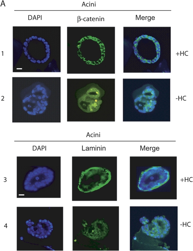

Generation of acini requires glucocorticoids. (A) Fluorescence microscopic analysis of 4 d acini stained with DAPI (left), anti–β-catenin antibody (middle) and merged image (right) cultured from time of seeding in the presence (+HC; row 1) and absence (−HC; row 2) of hydrocortisone (all 40X). Bar, 10 μm. Rows 3 and 4, same as above but stained with DAPI (left), anti–laminin V antibody (middle), and merged image (right). (B) RT-PCR analysis of total RNA isolated from 4 d acini cultured in the presence and absence of hydrocortisone for cyclin D1 and p21 transcripts (i); and 2 and 4 d acini and 2 d acini cultured in the absence of hydrocortisone (2d-HC) for occludin, ZO1, ZO2, and ZO3 transcripts (ii). GAPDH mRNA levels were measured as a control for equal input amounts of RNA. No reverse transcriptase (RT) added controls, lanes (−), show absence of DNA contamination. (iii) Western analyses of ZO1 and occludin in 2 and 4 d acini and in acini cultured in the presence of RU486 (50 μM; RU). β-Actin was measured as a loading control. (C) EMSA analysis, using a 32P-labeled NFκB-DNA binding element, performed on whole cell extracts from mammary epithelial cells at harvest (H, lane 1), cells maintained as monolayers on plastic (M, lane 2), cells cultured on concentrated EHS ECM (acini) for 2 and 4 d (lanes 3 and 4), monolayers on dilute EHS ECM matrix (lanes 5 and 6), monolayers on plastic overlaid with the EHS ECM matrix (lanes 7 and 8), and acini cultured in the presence of (+) and absence of (−HC) hydrocortisone (lanes 9 and 10). Arrowheads indicate NFκB complexes. PNFκB indicates binding element. In this and subsequent figures all discontinuities in gel images are marked with a vertical line. (D) EMSA analysis, as C, but using 32P-labeled AP1-DNA binding element. Light colored arrowheads indicate acinus-associated complexes. (E) Western analysis of phospho(ser63)-c-Jun (i), total c-Jun (ii), IP (JunD)/Western (anti–phospho-serine) (iii), and IP (JunD)/Western (JunD) (iv), in 2 and 4 d acini and in acini cultured in the presence of RU486 (50 μM; RU).