
Figure 4.
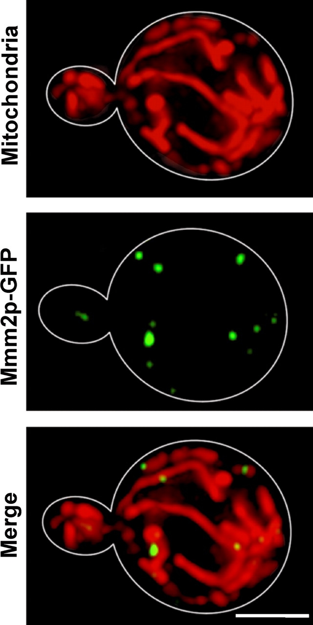
Mmm2p-GFP localizes to small spots along mitochondrial tubules. mmm2Δ strain RJ713, expressing the Mmm2p-GFP fusion protein from pAAH13, was stained with Mitotracker Red and examined by deconvolution microscopy. A total of 15 images were taken at 0.2-μm intervals in the z-axis, deconvolved, and then compressed into a single image. Mitotracker Red (Mitochondria), Mmm2p-GFP, and merged images with both Mmm2p-GFP and Mitotracker Red are shown. Bar, 3 μm.