
Figure 7.
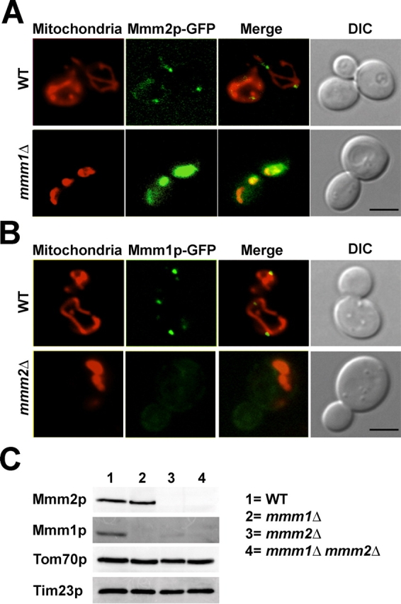
Mmm1p is necessary for the punctate distribution of Mmm2p, while Mmm2p is required for normal Mmm1p levels. (A) Wild-type strain MY4 and mmm1Δ strain MY5, each expressing Mmm2p-GFP from chromosomal MMM2::GFP, were stained with Mitofluor 589 and examined by DIC and fluorescence microscopy. Mitofluor 589 (Mitochondria), Mmm2p-GFP, merged images with both Mmm2p-GFP and Mitofluor 589, and DIC images are shown. Bar, 3 μm. (B) Wild-type strain SS12 and mmm2Δ strain AAH4, each carrying a chromosomal version of MMM1::GFP, were stained with Mitofluor 589 and examined by DIC and fluorescence microscopy. Mitofluor 589 (Mitochondria), Mmm1p-GFP, merged images with both Mmm1p-GFP and Mitofluor 589, and DIC images are shown. Bar, 3 μm. (C) Mitochondria (100 μg) were isolated from isogenic wild-type, mmm1Δ, mmm2Δ, and mmm1Δ mmm2Δ cells and Western blotted using antibodies to Mmm2p, Mmm1p, Tom70p, an outer membrane protein, and Tim23p, an inner membrane protein.