
Figure 1.
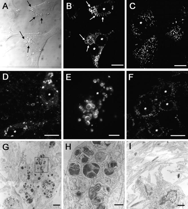
Enlarged lysosomal organelles are visible in PS1 −/− cells. (A) Differential contrast interference imaging of the cell surface of PS1 −/− fibroblasts demonstrated that a significant volume of the cells was occupied by large, spherical structures (arrows point to examples of enlarged vesicular structures). Lamp-2 immunolabeling of PS1 −/− (B) or PS1 +/? (C) fibroblasts demonstrated that these enlarged structures expressed lysosomal marker proteins and were present only within PS1 −/− cells. Similar accumulations of Lamp-2–positive organelles were detected in (D and E) PS1 −/− but not (F) PS1 +/? mouse primary neurons. Transmission EM analysis of PS1 −/− (G and H) or PS1 +/?. (I) Neurons demonstrated that PS1 −/− neurons contain large clusters of organelles. H is a higher magnification of the boxed area in G. *, denotes nucleus. Bars: (B–D and F) 10 μm; (E) 5 μm; (G and I) 1 μm; (H) 200 nm.