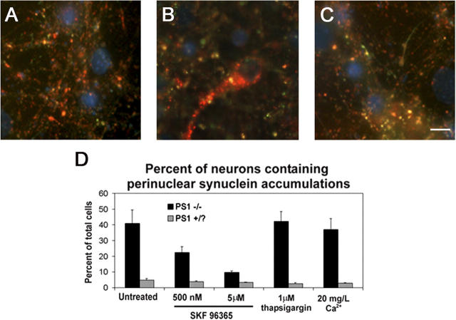
Figure 9.
Inhibition of capacitative calcium channels eliminates the perinuclear distribution of synucleins in PS1 −/− neurons. PS1 −/− and PS1 +/? neurons cultured for 10 d in vitro were treated with SKF 96365 or thapsigargin, or incubated in low Ca2+ medium for 48 h. Representative images of (A) untreated PS1 +/? neurons, (B) untreated PS1 −/−neurons, or (C) PS1 −/− neurons treated with 500 nM SKF 96365 are shown. Nuclei are counterstained with DAPI (blue). Bar, 10 μm. (D) Quantification of the percentage of neurons displaying perinuclear synuclein immunoreactivity. Error bars represent SD.

