
Figure 4.
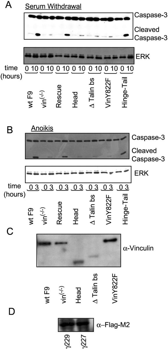
Role of various regions of vinculin in the control of apoptosis induction in a different F9 vin − / − clone (γ227). (A) Cell extracts from WT F9 and F9 vin−/− cells (γ227 clone) stably expressing each construct were analyzed by immunoblotting using anti–caspase-3 Ab 10 h after serum withdrawal. (B) Immunoblotting analysis of cell extracts from WT F9 and F9 vin−/− cells stably expressing each construct 3 h after disruption of cell–matrix interaction using anti–caspase-3 Ab. The results shown in A and B are representative examples from two independent experiments. (C) Expression of full-length vinculin, head, Δ talin bs, and vinY822F in γ227 clones. (D) Expression of the Flag M2–tagged hinge-tail fragment in γ229 and γ227 clones.