Figure 1.

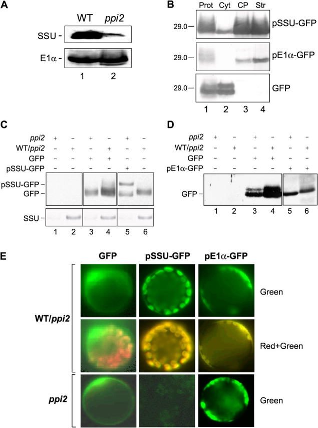
In vivo targeting of transit peptide–GFP fusion proteins to plastids in heterozygous or homozygous ppi2 seedlings. Heterozygous ppi2 plants were transformed with binary vector constructs encoding pSSU-GFP, pE1 α-GFP, or GFP. (A) Levels of endogenous SSU and E1α in wild-type (WT) and ppi2 plants. Extracts from 3-wk-old plants (75 μg protein) were resolved by SDS-PAGE and immunoblotted with antisera to each protein. (B) Enrichment of pSSU-GFP and pE1α-GFP in chloroplast fractions. Protoplasts (Prot) from heterozygous ppi2 plants expressing either pSSU-GFP, pE1α-GFP, or GFP alone were fractionated into cytoplasmic (Cyt), total chloroplast (CP), and chloroplast stroma (Str) fractions. The fractions (12 μg protein) were immunoblotted with anti-GFP antibodies. The 29-kD marker is indicated to the left of each panel. (C and D) Immunoblot analysis of total protein extracts from heterozygous ppi2 (WT/ppi2) and homozygous ppi2 (ppi2) plants expressing GFP, pSSU-GFP, or pE1α-GFP with anti-GFP antibodies (top panels) or with an anti-SSU serum (C, bottom). Lanes 1 and 2 (C and D) contain protein extracts from plants not transformed with GFP constructs. Black lines indicate grouping of images from different portions of the same gel. Images from different gels are in separate boxes. (E) Confocal laser scanning microscopy of protoplasts isolated from heterozygous ppi2 (WT/ppi2) or homozygous ppi2 (ppi2) plants expressing GFP, pSSU-GFP, or pE1α-GFP. GFP fluorescence (Green) and the merge of chlorophyll autofluorescence and GFP fluorescence (Red + Green) are shown.
