
Figure 1.
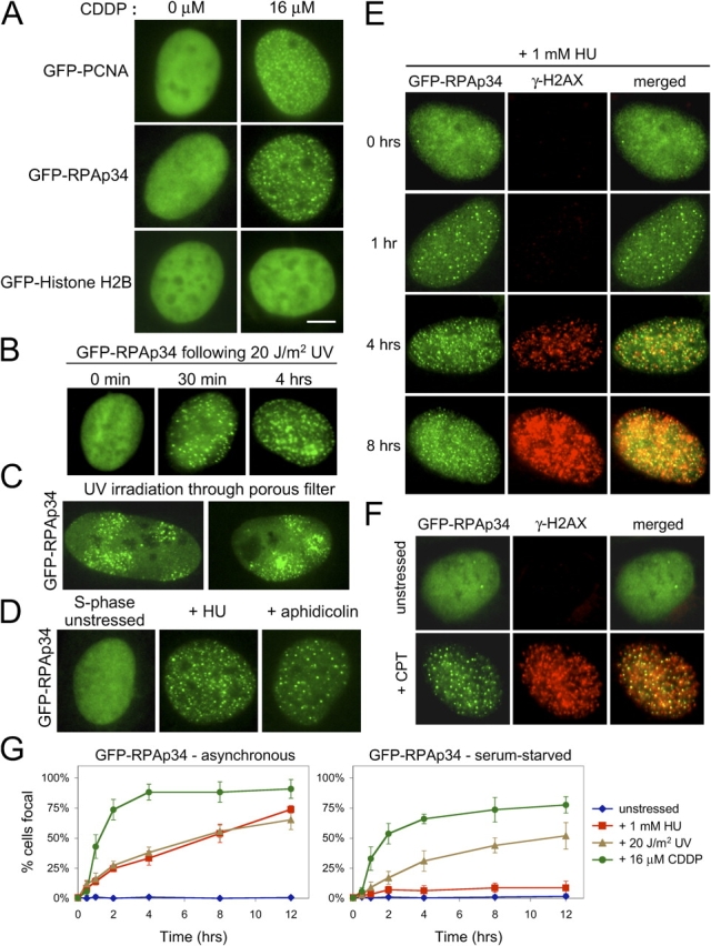
Visualization of replication factors in the DNA damage response. (A) CDDP induces the relocalization of GFP-PCNA and GFP-RPAp34, but not GFP-histone H2B, into discrete foci within the nucleus of stably expressing Rat-1 cells. Bar, 5 μm. (B) RPAp34 rapidly accumulates into foci in response to UV irradiation. (C) Cells expressing GFP-RPAp34 were exposed to UV irradiation through a porous polycarbonate filter. GFP-RPAp34 only accumulates into foci within the irradiated microdomains 3 μm in diameter. (D) RPAp34 accumulates into foci in response to the stalling of DNA replication with HU or APH. (E) RPAp34 foci appear before the accumulation of H2AX phosphorylation after HU treatment. (F) Camptothecin (CPT) induces the formation of RPAp34 foci that colocalize with γ-H2AX foci. (G) Quantitation of the number of cells with punctate versus diffuse localization of GFP-RPAp34 in asynchronous (left) or serum-starved (right) cultures after various stresses over time. 200 cells were counted at each time point from each of three independent experiments. After 48 h serum-starvation, 4% of the unstressed cells labeled BrdU positive during the 12-h time course of the experiment.