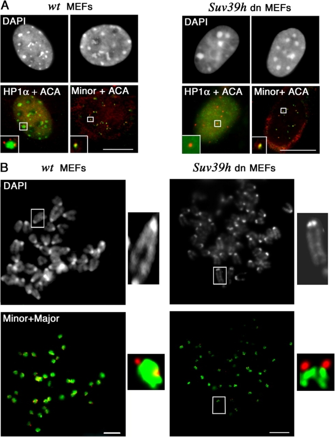
Figure 7.

Major and minor satellite domains in Suv39h double mutant cells. (A) Major and minor satellite chromatin in MEFs Suv39h double mutant (Suv39h dn) compared with wild-type (wt) nuclei. Costaining with ACA serum (red) and anti-HP1α antibodies (green). ACA immunostaining (red) combined with minor satellite FISH (green). DNA was visualized with DAPI. Insets correspond to close-ups of selected foci. Bars, 5 μm. (B) Sister chromatid separation of minor and major satellites at metaphase stage in MEFs Suv39h double mutant (Suv39h dn) compared with wild type (wt). DNA FISH for major (green) and minor (red) satellites. DNA was visualized with DAPI. A close-up of a selected chromosome is shown along each panel (insets). Bars, 2 μm.
