
Figure 1.
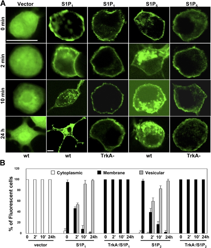
NGF-induced S1P receptor internalization is TrkA dependent. (A and B) Naive or TrkA-less nnr5 PC12 cells were transfected with expression plasmids encoding GFP, S1P1-GFP, or S1P5-GFP and were cultured for 48 h. Serum-starved cells were treated with 100 ng/ml NGF for the indicated times and were visualized by confocal microscopy. (A) Representative cells of more than 100 cells examined are shown. (B) S1P receptor localization was quantified by assessing the percentage of GFP-positive cells with cytoplasmic staining (open bars, GFP was present inside the cell without any indication of discrete structural staining), membrane staining (black bars, GFP outlined the cell with little if any GFP present inside the cell), or vesicular staining (gray bars, GFP was present inside the cell in discrete, punctate patterns). The absence of bars indicates none detected. Results are means ± SD from four independent experiments. At least 100 cells per condition were counted. Bars, 25 μm.