
Figure 3.
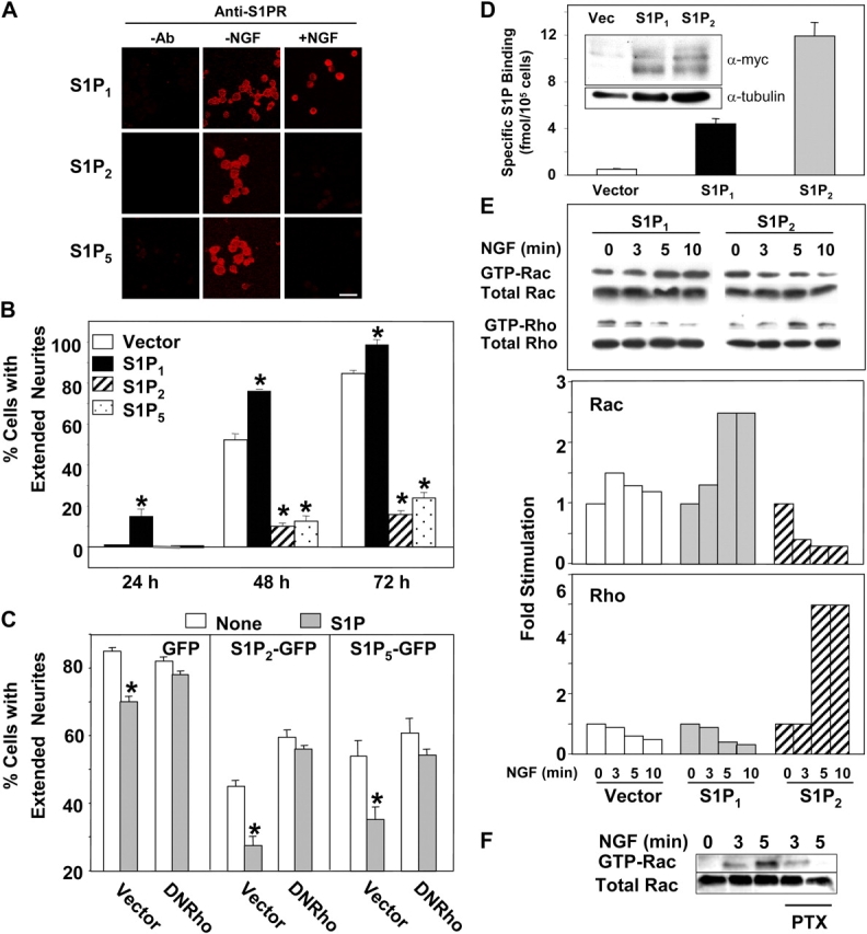
Activation of S1P1 stimulates, whereas S1P2 and S1P5 inhibit neurite extension. (A) PC12 cells were plated on poly-d-lysine–coated coverslips and cultured in the absence (−) or presence (+) of NGF (100 ng/ml) for 24 h. Immunocytochemical analysis of S1P receptor expression was performed with specific antibodies as indicated and was visualized by rhodamine-conjugated secondary antibody. No appreciable background staining was evident with IgG or when primary antibody was omitted or preincubated with the peptide antigen. Bar, 100 μm. (B) PC12 cells were transfected with vector-GFP (open bars), S1P1-GFP (filled bars), S1P2-GFP (hatched bars), or S1P5-GFP (stippled bars) and were cultured for 48 h. Serum-starved cells were treated with 100 ng/ml NGF for the indicated times and neurite extension was quantified by assessing the percentage of GFP-positive cells bearing at least one neurite twice the length of the cell body. Asterisks denote significant differences relative to vector transfectants (P < 0.01, ANOVA, Tukey's). (C) PC12 cells were cotransfected with vector or N19RhoA (DNRho) together with vector-GFP, S1P2-GFP, or S1P5-GFP, as indicated, at a ratio of 5:1 to ensure that GFP-expressing cells also express DNRho. Serum-starved cells were incubated with 100 ng/ml NGF for 120 h and treated without (open bars) or with S1P (100 nM; gray bars) for the last 3 h. Neurite extension was quantified by assessing the percentage of GFP-positive cells bearing at least one neurite twice the length of the cell body. Asterisks denote significant differences relative to untreated cells (P < 0.01, ANOVA, Tukey's). (D) Specific binding of [32P]S1P (defined as binding in the absence of unlabeled competitor minus binding in the presence of excess unlabeled ligand) to PC12 cells stably expressing vector, myc-S1P1, or myc-S1P2 was measured as described previously (Van Brocklyn et al., 1999). Inset: Western blot of cell lysates was probed with anti-Myc or anti-tubulin antibodies (Santa Cruz Biotechnology, Inc.) as described previously (Watterson et al., 2002). (E) Serum-starved PC12 cells stably transfected with vector (open bars), myc-S1P1 (gray bars), or myc-S1P2 (hatched bars) were stimulated with 100 ng/ml NGF for the indicated times. Activated Rac and Rho were specifically pulled down from cell lysates containing equal amounts of proteins and were analyzed by Western blotting using anti-Rac and anti-Rho antibodies, respectively. Relative activated levels were normalized to total Rac and Rho and the fold stimulation is depicted. Representative results from one of three independent experiments are shown. (F) Wild-type PC12 cells were cultured in serum-free medium without or with PTX (100 ng/ml) for 16 h, then stimulated with NGF for the indicated times and Rac activation determined.