
Figure 6.
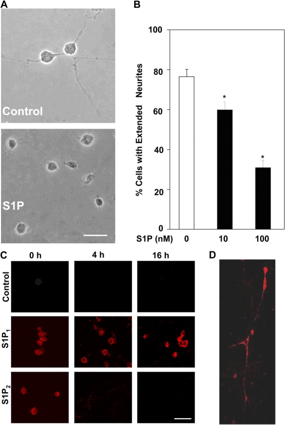
S1P suppresses NGF-induced neurite extension in freshly dissociated DRG neurons. (A) Rat E15 DRG neurons were dissociated, plated on growth factor–reduced Matrigel™-coated coverslips and treated without or with 100 nM S1P (A) or the indicated concentrations of S1P (B) in 100 ng/ml NGF-containing medium. Photomicrographs of representative neurons examined by phase microscopy are shown. (B) Neurite extension was quantified after 16 h by assessing the percentage of neurons bearing at least one neurite four times the length of the cell body. A minimum of 300 neurons was examined and data are the averages ± SD. Similar results were obtained in three independent experiments. Asterisks denote significant differences relative to untreated cells (P < 0.01, ANOVA, Tukey's). (C) Differential expression of S1P receptors during development of DRG neurons. Rat E15 DRG neurons were dissociated, plated on growth factor–reduced Matrigel™-coated coverslips, and cultured in the presence of NGF for 0, 4, and 16 h. Immunocytochemical analysis of S1P receptor expression was performed with specific antibodies as indicated and stained with rhodamine-conjugated secondary antibody and visualized by fluorescence microscopy. No appreciable background staining was evident with IgG or when primary antibody (control) was omitted or preincubated with the peptide antigen. (D) Confocal collage of a representative neuron treated with NGF for 16 h and stained with anti-S1P1 showing expression of S1P1 on extended neurites. Bars: 50 μm (A); 100 μm (C).