Figure 8.
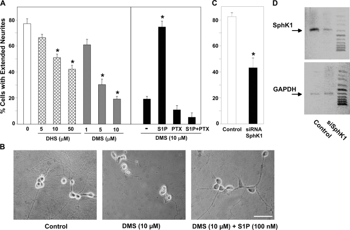
SphK1 activity is required for neurite extension. (A) Rat E15 DRG neurons were dissociated, plated on growth factor–reduced Matrigel™-coated coverslips, and treated with the indicated concentrations of d,l -erythro-dihydrosphingosine (DHS, hatched bars) or N,N-dimethylsphingosine (DMS, gray bars), and were cultured for 16 h in the presence of NGF. Parallel cultures (right) incubated with 10 μM DMS (black bars) were subsequently treated with 200 ng/ml PTX for the final 3 h, 100 nM S1P for the final 1.5 h, or both. Neurite extension was quantified. (B) Photomicrographs of representative neurons examined by phase microscopy are shown. Note that S1P rescued the DMS-inhibited neurite extension. Bar, 100 μm. (C) Rat E15 DRG neurons were dissociated and transfected with a control siRNA sequence or with siRNA targeted to SphK1. After 120 h in the presence of NGF, neurite outgrowth was quantified by assessing the percentage of neurons bearing neurites four times the length of their cell bodies. Asterisks in A and C denote significant differences relative to untreated cells (P < 0.01, ANOVA, Tukey's). (D) RT-PCR analysis of SphK1 and GAPDH expression in DRGs transfected with control siRNA or siSphK1.

