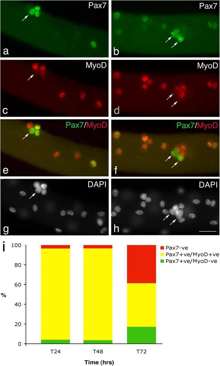
Figure 1.

Satellite cells adopt divergent fates. After 72 h in culture, coimmunostaining of EDL myofibers demonstrates that in the majority of satellite cells, Pax7 (a and b) is becoming down-regulated whereas MyoD (c and d) remains strongly expressed. However, combining the Pax7 (green) and MyoD (red) fluorescent immunosignals (e and f) clearly shows that some (∼23%) MyoD−ve daughters (arrows) maintain robust Pax7 expression. Pax7+ve/MyoD−ve satellite cell (arrows) are typically located (85%) in clusters (defined as four or more overlying/touching cells) together with both Pax7+ve/MyoD+ve and Pax7−ve/MyoD+ve cells. Counterstaining with DAPI was used to identify all nuclei present on the myofiber (g and h). Bar, 30 μm. The number of satellite cells from 3F-nlacZ-E mouse 3 and 4 (Table II) that were either Pax7+ve/MyoD−ve, Pax7+ve/MyoD+ve, or Pax7−ve at T24, T48, and T72 were counted. Expressing this data as the mean percentage in each category of the total number of satellite cells per myofiber (i) illustrates that the Pax7+ve/MyoD+ve cells present at T24 and T48 give rise to the majority of Pax7+ve/MyoD−ve and Pax7−ve populations at T72.
