
Figure 3.
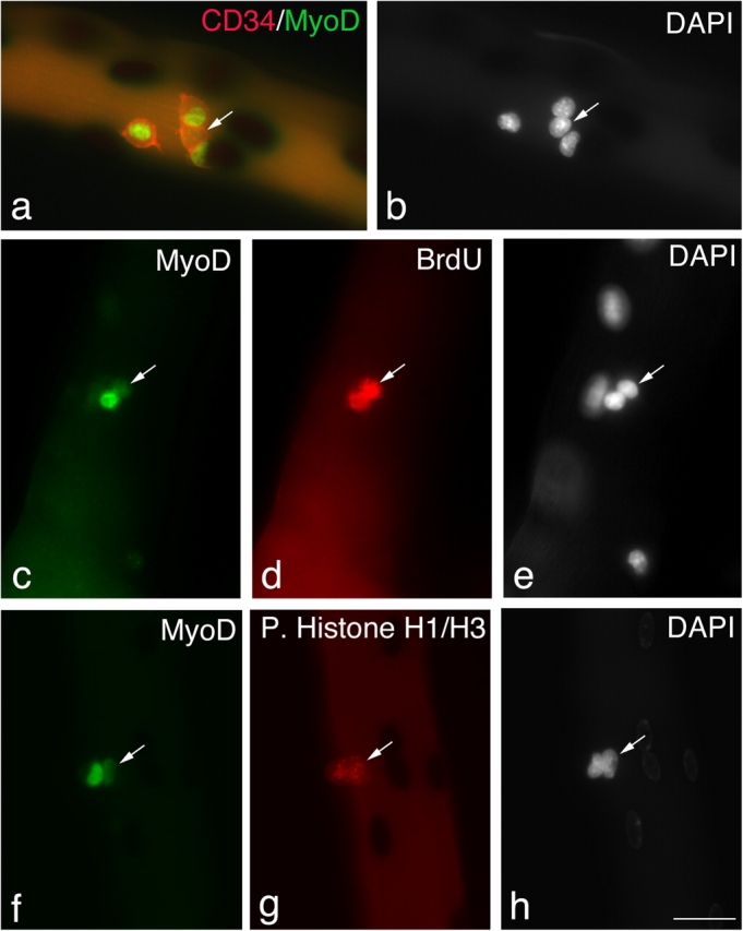
Satellite cells can maintain Pax7, but lose MyoD. Quiescent satellite cells are generally distributed as single cells along the length of the myofiber. Between 24–48 h in culture, almost all satellite cells express both Pax7 and MyoD and begin to divide; therefore, it can be assumed that most pairs/small groups of cells present at these times are derived from a single parent satellite cell. Immunostaining followed by X-gal incubation of EDL myofibers from a 3F-nlacZ-E mouse after 48 h in culture shows that of four CD34+ve satellite cells (a, red-cell surface), three contained MyoD protein (a, green nuclear), but significantly one did not (a and b, arrows). These MyoD−ve cells (c–e, arrows) had undergone division, as shown by the incorporation of BrdU into the daughter cells (d), and were not merely quiescent satellite cells. Whether MyoD protein levels vary with cell cycle is unresolved (Kitzmann et al., 1998; Lindon et al., 1998). However, it is important that both the MyoD+ve and MyoD−ve progeny of a single satellite cell could be in the same phase of the cell cycle (f–h). An example is shown where immunostaining followed by X-gal incubation of an EDL myofiber from a 3F-nlacZ-E mouse after 48 h in culture shows that MyoD+ve (f) and MyoD−ve (f–h, arrows) daughters are both in the same phase of the cell cycle as shown by phosphorylated Histone H1/H3 immunostaining (g). Counterstaining with DAPI was used to identify all nuclei present (b, e, and h). Bar, 30 μm.