
Figure 8.
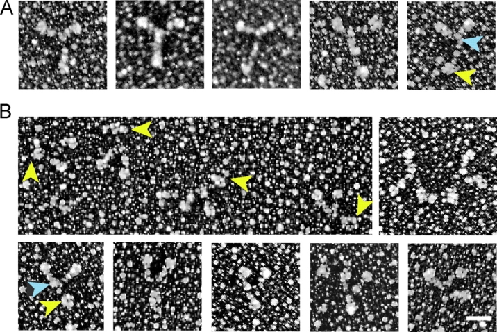
Metal-shadowed images of full-length myosin V. Myosin V in (A) an extended or (B) more compact conformation. Yellow arrowheads point to the globular tail, and blue arrowheads to the head–rod junction. Note that the length of the tail is shorter in the compact conformation. The molecules shown in A were diluted into high ionic strength for rotary shadowing, whereas those in B were obtained in a lower ionic strength buffer. In rows 1 and 3, the molecules are oriented with the heads up and the tail down. Bar, 30 nm.