Figure 1.
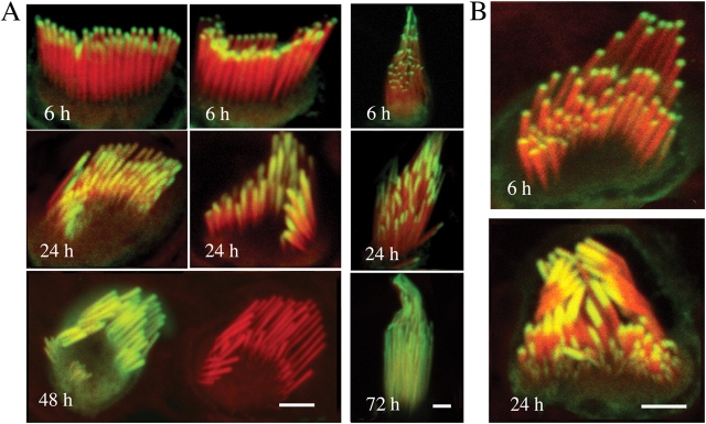
Incorporation of β actin and espin in stereocilia. (A) β actin–GFP incorporation into the stereocilia of the hair cells of organ of Corti (left) and vestibular organs (right) after transfection. Confocal microscopy revealed that β actin–GFP (green) appeared at the tips of stereocilia counterstained with rhodamine/phalloidin (red) 6 h after transfection. β actin–GFP was progressively incorporated from the stereocilia tips to their bases within 48 h in the organ of Corti hair cells and within 72 h in the vestibular hair cells. Stereocilia maintained their lengths as evident in the bottom left panel where actin-GFP fluorescence had reached the base of the stereocilia, yet the stereocilia length is similar to the neighboring nontransfected cell. Bars, 2.5 μm. (B) Espin-GFP incorporation into the stereocilia of the hair cells of organ of Corti. Espin-GFP incorporation began at the tips of stereocilia and was progressively incorporated into stereocilia at similar rates as the β actin–GFP shown in A. Bar, 2 μm.

