
Figure 5.
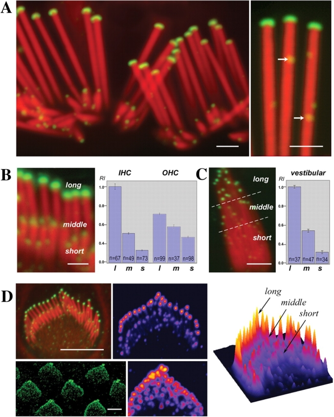
Intensity of myosin XVa labeling in the tips of stereocilia is graded with length. (A–C) Myosin XVa is localized to the stereocilia tips of auditory (A and B) and vestibular (C) hair cells. Confocal images revealed myosin XVa immunofluorescence in all stereocilia tips of the rat auditory hair cells (A) and at higher magnification also exhibited small fluorescence puncta (A, right, arrows) along the entire stereocilia. Levels of myosin XVa labeling are highest in the longest stereocilia within the auditory (B) and vestibular (C) hair bundle. The relationship between myosin XVa and stereocilia length is visible in fully developed bundles (A) as well as in cultured auditory (B) and vestibular hair cells (C). Quantitative analysis of average pixel intensity confirms myosin XVa gradation within hair bundles of inner (IHC) and outer (OHC) auditory hair cells (B) as well as vestibular hair cells (C). Error bars equal mean ± standard error of the mean. l, long; m, middle; s, short stereocilia. The gradient of myosin XVa expression is noticeable at the earliest stages of the staircase formation in inner (D, top) and outer (D, bottom) hair cells from postnatal day 1 rat neonates whose shorter stereocilia are indistinguishable from supernumerary microvilli. Scaling of myosin XVa levels to the stereocilia length and their distribution within a bundle is clearly visible on the pseudo-colorized images and surface plot of pixel intensities (D); the highest intensities are shown in red. Bars: (A and B) 1 μm; (C) 2 μm; (D) 4 μm.