
Figure 6.
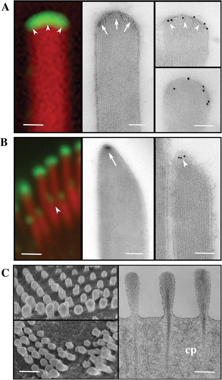
Myosin XVa is a component of the stereocilia tip complex. (A and B) Comparison of high magnification confocal immunofluorescence (left), TEM of uranyl acetate–stained sections (middle), and immunogold-labeled unstained sections (right) of the oblate (A) and prolate (B) stereocilia tips. Stereocilia tips of the tallest row of the bundle are prolate in shape with an electron-dense region just below the stereocilia membrane (A, arrows), where intense myosin XVa labeling is visualized by immunofluorescence or by immunogold EM (A, arrowheads). The second row stereocilia in the bundle are prolate and pointed (B) with a smaller discrete electron-dense region at the end of their longest actin filaments (B, arrow), which also coincides to where myosin XVa is visualized by either fluorescence or by immunogold EM (B, arrowheads). (C) SEM images of shaker 2J mutants confirm previous observations that stereocilia, despite being very short, maintain a slight length gradation reminiscent of the staircase pattern of a normal hair bundle. TEM of osmium and uranyl acetate–stained thin sections (right) show that these very short stereocilia maintain the overall organization of the actin filaments including the rootlets that insert into the cuticular plate (cp) but their tips lack the electron-dense material seen in normal stereocilia (A and B). Bars: (A) 100 nm; (B) 1 μm (left) and 100 nm (right); (C) 1 μm (left) and 100 nm (right).