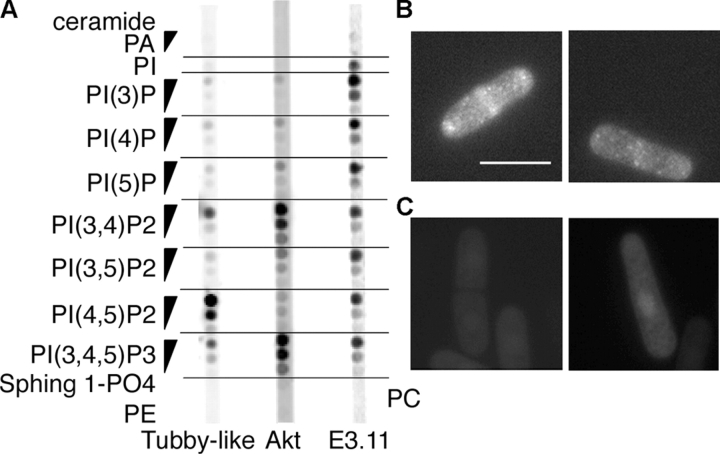
Figure 6.
Phosphoinositide-binding PH domain regulated by ptn1p. (A) Binding of 35S-labeled PH domains to lipids immobilized on a membrane. The Tubby-like and Akt PH domains show specificity for PI(4,5)P2 and PI(3,4)P2+PI(3,4,5)P3, respectively. The 11E3.11C PH domain from S. pombe binds many phosphoinositides, including PI(3,4,5)P3. (B) Distribution of GFP-11E3.11C PH domain in ptn1Δ cells. The left and right panels show a dividing cell and a growing cell, respectively. Bar, 10 μm. (C) GFP-11E3.11C PH domain shows no clear localization in wild-type cells.

