
Figure 2.
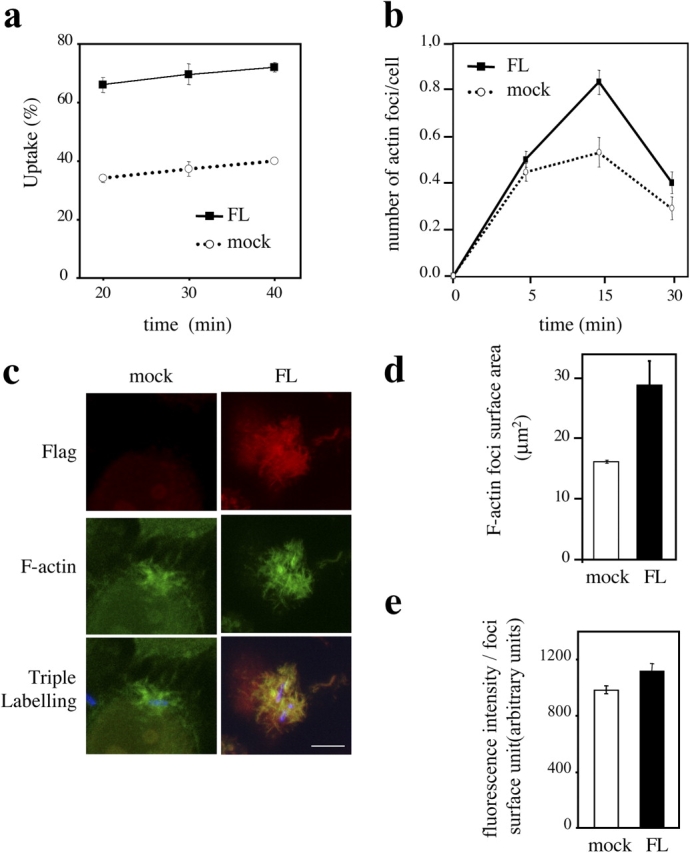
Overexpression of cortactin enhances Shigella-induced actin polymerization and bacterial uptake. HeLa cells were transfected with vector alone (mock) or Flag-tagged cortactin (FL). After 24 h, cells were infected with Shigella, fixed, and processed for fluorescent labeling. Transfected cells were visualized by staining of FL with anti-Flag antibody. (a) Shigella uptake is shown for mock cells (dotted line) and FL cells (solid line). (b) The frequency of Shigella-induced actin foci formation is shown for mock cells (dotted line) and FL cells (solid line). (c) Representative confocal images of Shigella foci observed in mock or FL cells, stained for Flag-tagged cortactin (red), F-actin (green), and Shigella (blue). Bar, 5 μm. (d and e) Quantitative analysis of the surface area and the fluorescence intensity per surface unit of F-actin foci was performed in mock cells (white bar) and FL cells (solid bar), using a dedicated computer program. All the plotted data shown in this figure were averaged from three independent experiments ± SEM.