
Figure 3.
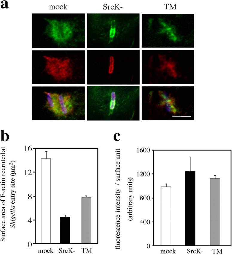
Expression of kinase-inactive Src and TM cortactin inhibits Shigella-induced actin foci formation. (a) Representative confocal microscopy images of Shigella entry structures observed in mock cells (mock), SrcK− cells (SrcK−), and cells expressing tyrosine-mutated cortactin (TM). Cells were challenged for 15 min with Shigella, fixed, and processed for fluorescence labeling of F-actin (green), cortactin (red), and bacterial LPS (blue). Bar, 5 μm. (b and c) Surface area and fluorescence intensity per surface unit of F-actin in Shigella entry structures were quantified in mock, SrcK−, and TM cells from images obtained with an epifluorescence microscope using a dedicated computer program (see Materials and methods). The analysis was performed on 60 foci from three independent experiments.