
Figure 9.
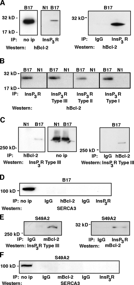
Coimmunoprecipitation of Bcl-2 with InsP3Rs. (A) InsP3Rs were immunoprecipitated from N1 and B17 clones using a subtype nonspecific antibody. Immunoprecipitates were analyzed by Western blotting using an antibody to hBcl-2. The level of overexpressed human Bcl-2 is shown by Western blotting in the “no ip” lane. (B) InsP3Rs were immunoprecipitated from N1 and B17 cells using antibodies that recognize either all three subtypes (left) or antibodies specific for each subtype (second through fourth panels). Immunoprecipitates were analyzed by Western blotting using anti–human Bcl-2 antibody. (C) Human Bcl-2 (i.e., overexpressed Bcl-2) was immunoprecipitated, and immunoprecipitates were analyzed by Western blotting using antibody specific for InsP3R subtype III. The levels of type III InsP3Rs are shown by Western blotting in the “no ip” lane. (D) Bcl-2 and InsP3Rs were immunoprecipitated from the B17 clone as described in preceding panels, and immunoprecipitates were analyzed by Western blotting using antibody to SERCA3. The level of SERCA3 in B17 cells is shown in the “no ip” lane. (E) Endogenous mouse Bcl-2 and InsP3Rs were immunoprecipitated from S49.A2 cells using an antibody specific for mouse Bcl-2 and an antibody that reacts with all three subtypes of InsP3Rs, respectively. In all panels, the lanes labeled “IgG” represent immunoprecipitations performed using nonspecific IgG control antibodies. InsP3Rs were detected in Bcl-2 immunoprecipitates using antibody to Type III InsP3Rs (left), while endogenous Bcl-2 was detected in InsP3R immunoprecipitates by probing Western blots with an antibody to murine Bcl-2 (right). (F) Bcl-2 and InsP3Rs were immunoprecipitated from S49A2 cells as described in preceding panels, and immunoprecipitates were analyzed by Western blotting using antibody to SERCA3. The level of SERCA3 in S49A2 cells is shown in the “no ip” lane.