
Figure 5.
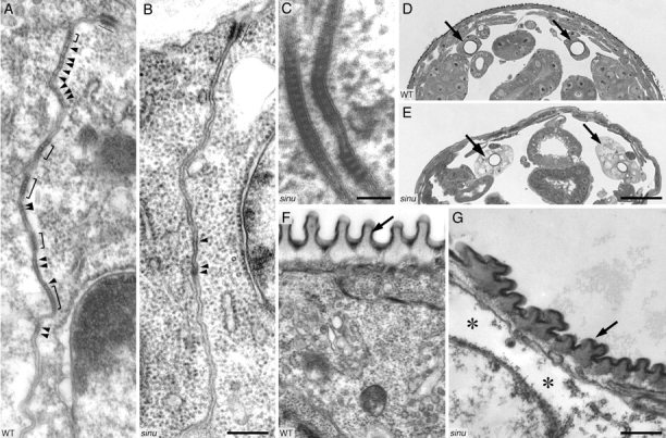
Sinuous is required for septate junction organization, but not for septa formation. (A) TEM of late stage 17 wild-type epidermis showing the zonula adherens (thin line) and septate junctions. Brackets indicate clustered groups of septa, and arrowheads point to individual septa. (B and C) TEM of late stage 17 sinuous mutant epidermis. In some sections, such as the one shown in B, septa are absent except for some poorly differentiated intercellular material (arrowheads) at cell–cell contacts. However, in other sections, such as the one shown in C, cell–cell contacts display normally organized septate junctions. The zonula adherens (B, thin line) appears normal in sinuous mutants. (D and E) Toluidine blue/methylene blue–stained cross sections of a stage 17 wild-type (D) and a sinuous mutant (E) embryo. Arrows point to the tracheal dorsal trunks. Note that the staining intensity of sinuous mutant tracheal cells is lighter and that the cell shape is more cuboidal than in the wild-type, whereas the epidermis in sinuous mutants stains similarly to wild-type epidermis. (F and G) TEM of dorsal tracheal trunks of late stage 17 wild-type (F) and sinuous mutant (G) embryos. In sinuous mutants, the taenidium (arrows) shows an irregular morphology, and the cytoplasm has large empty spaces (asterisks). Genotypes: WT, Oregon-R; sinu nwu7. Bars: B, 200 nm (for A and B); C, 50 nm; E, 25 μm (for D and E); G, 300 nm (for F and G).