Figure 5.

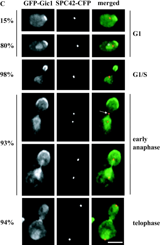
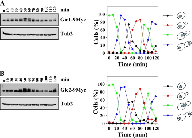
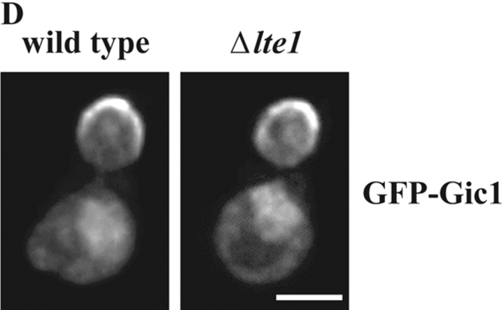
Gic1 was released from the bud cortex during anaphase. (A) Gic1 protein levels remain constant throughout the cell cycle. Gal1-CDC20 GIC1-9Myc cells were arrested in metaphase by incubating cells in YP raffinose medium (no induction of Gal1-CDC20). Galactose was added (t = 0) to the synchronized cells to induce CDC20 expression and to trigger anaphase onset. Samples were taken at the indicated time points after galactose addition. Cells were fixed, stained with DAPI and analyzed by fluorescence microscopy (n > 100). The circles in the cartoon cells indicate the DAPI staining regions. In addition, cell extracts were analyzed by immunoblotting with anti-Myc and anti-Tub2 (loading control) antibodies. (B) Gic2 is expressed in a cell cycle–dependent manner. Gal1-CDC20 GIC2-9Myc cells were treated and analyzed as in A. (C) Gic1 is localized to the bud cortex and the nucleus. Localization of Gic1 was determined using SPC42-CFP Gal1-GFP-GIC1 cells. Gal1-GFP-GIC1 of logarithmically growing cells was induced for 1 h by the addition of galactose. Fixed cells were analyzed by deconvolution fluorescence microscopy. The percentages indicate the frequencies of the various cell types at different stages of the cell cycle. In the remaining cells GFP-Gic1 did not show any specific cellular distribution. The asterisk indicates nuclear GFP-Gic1. The arrow points toward a cell in anaphase in which the SPB in the bud is exposed to Gic1. (D) Bud cortex association of Gic1 is independent of LTE1. Gal1-GFP-GIC1 of logarithmically growing LTE1 and Δlte1 cells was induced for 1 h by the addition of galactose. In most (98%) early anaphase cells GFP-Gic1 was associated with the bud cortex. Bars, 5 μm.


