
Figure 1.
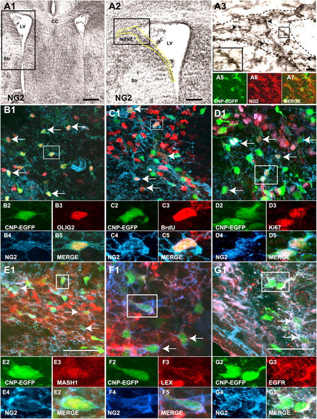
NG2 + /EGFP + cells express NSCs markers in the SVZ. Coronal sections of the SVZ at P8. (A1–A3) Anti-NG2 staining (DAB reaction, brown) shows that NG2 cells are found lining the wall of the lateral ventricle (yellow line) and throughout the entire lateral SVZ. The dotted line (A3) indicates the area analyzed in this paper. (A5–A7) Most of the EGFP+ cells (A5, green) were labeled with NG2 antibodies (A6, red), and all NG2+ cells were EGFP+ (A7). (B–G) All micrographs were obtained from the lateral SVZ (laSVZ). (B) All NG2+/EGFP+ cells (blue/green, respectively) express Olig2 (red). (C and D) NG2+/EGFP+ cells proliferate in the SVZ, as shown by BrdU incorporation (C, red) and by Ki67 immunolabeling (D, red). (E and G) A large percentage of NG2+/EGFP+ cells (blue/green, respectively) express the transcription factor Mash1 (E, red), the adult NSC markers LeX antigen (F, red), and EGFR (G, red). Arrows indicate NG2+/EGFP+ cells double-labeled with any of the markers used. NG2+/EGFP+ cells in boxed areas are shown at higher magnification. LV, lateral ventricle; Str, striatum; CC, corpus callosum. Bars: (A1) 500 μm; (A2) 300 μm; (A3) 50 μm; (B–E) 50 μm; (F and G) 100 μm.