
Figure 1.
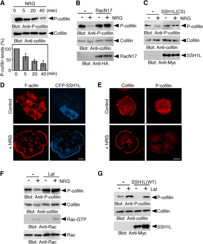
NRG stimulates cofilin dephosphorylation in a manner dependent on Rac, SSH1L, and actin assembly. (A) NRG induces cofilin dephosphorylation. MCF-7 cells were stimulated with NRG. Cell lysates were immunoblotted with antibodies against P-cofilin and cofilin. Relative P-cofilin levels are shown as means ± SEM of triplicate experiments. (B and C) Expression of RacN17 or SSH1L(CS) inhibits NRG-induced cofilin dephosphorylation. MCF-7 cells transfected with HA-RacN17 (B) or Myc-SSH1L(CS) (C) were stimulated with NRG for 20 min and the P-cofilin levels were analyzed as in A. (D) SSH1L accumulates in NRG-induced lamellipodia. MCF-7 cells expressing CFP-SSH1L were stimulated with NRG for 15 min and stained with rhodamine phalloidin for F-actin. Bar, 20 μm. (E) Cofilin, but not P-cofilin, accumulates in NRG-induced lamellipodia. MCF-7 cells were stimulated with NRG for 15 min and stained with antibodies against cofilin or P-cofilin. Bar, 20 μm. (F) Lat-A inhibits NRG-induced cofilin dephosphorylation. MCF-7 cells were treated with Lat-A for 10 min, then stimulated with NRG for 20 min, and the P-cofilin levels were analyzed. Active GTP-bound form of Rac was analyzed using pull-down assay with GST-PBD (p21-binding domain of PAK1; Nishita et al., 2002). (G) Lat-A inhibits SSH1L- induced cofilin dephosphorylation. MCF-7 cells transfected with Myc-SSH1L(WT) were treated with Lat-A for 30 min and the P-cofilin levels were analyzed as in A.