
Figure 1.
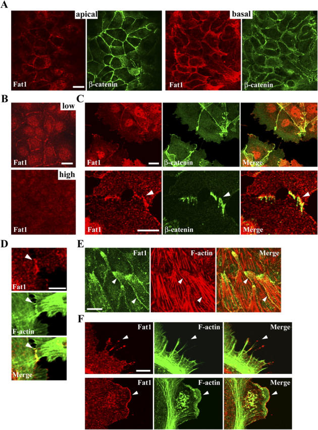
Subcellular localization of endogenous Fat1. (A) DLD1 cells doubly immunostained for Fat1 (red) and β-catenin (green). An apical and a basal confocal section are shown. Fat1 is localized at cell junctions, but is more abundant in the basal portion of the junctions. (B) MDCK cells immunostained for Fat1 at a low and high density. The junctional Fat1 tends to disappear at high cell densities. (C) PAM212 cells doubly immunostained for Fat1 (red) and β-catenin (green), showing localization at cell junctions (top panels). Bottom panels, close-up view of early cell–cell contacts. Fat1 is detected at cell contact sites, but its localization pattern is not identical to that of β-catenin (arrowhead). (D) Early contacts between PAM212 cells doubly immunostained for Fat1 (red) and F-actin (green). Fat1 colocalizes with junction-associated F-actin (arrowhead). (E) A confocal section of PAM212 cells at the basal level of the cell layer, doubly immunostained for Fat1 (green) and F-actin (red). Fat1 tends to accumulate at a protruded portion of cells as well as at cell–cell contacts (arrowhead), overlapping with F-actin. (F) Close-up views of the free surface of PAM212 cells doubly immunostained for Fat1 (red) and F-actin (green). Fat1 is localized to filopodia (top, arrowhead) and lamellipodia (bottom, arrowhead), overlapping with F-actin. Bars: 15 μm (A, B, and E), 10 μm (C, top), 5 μm (C, bottom; D and F).