
Figure 4.
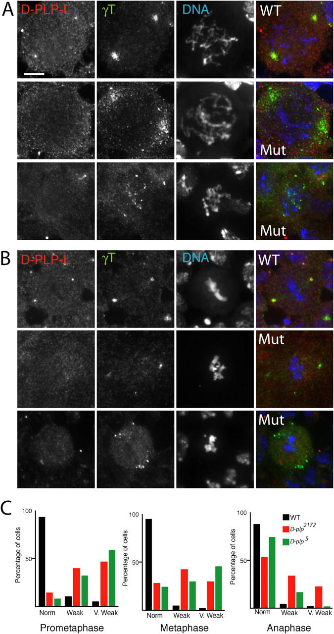
The mitotic recruitment of γ-tubulin to centrosomes is impaired in d-plp mutant cells. The distribution of D-PLP-L (left panels), γ-tubulin (middle panels), and DNA (right panels) is shown in WT and d-plp mutant larval brain cells in prometaphase (A) and metaphase (B). In WT prometaphase cells, γ-tubulin is recruited to a relatively broad area around the centrioles. In mutant prometaphase cells, D-PLP-L is no longer detectable at centrioles, and γ-tubulin is (1) often only weakly recruited to one or two diffuse areas (middle panels); or (2) remains scattered throughout the cell (bottom panels). In WT metaphase cells, γ-tubulin is recruited to a compact area around the centrioles. In mutant metaphase cells, the centrosomal recruitment of γ-tubulin is often weaker than normal (middle panels), and in some cells, particles of γ-tubulin remain scattered in the cytoplasm (bottom panels). Bar, 5 μm. (C) Quantitation of the γ-tubulin localization defects in WT and mutant brain cells. The graphs show the percentages of cells displaying normal, weak, or very weak centrosomal localization of γ-tubulin in WT (black), d-plp 2172 (red), or d-plp 5 (green) cells in prometaphase, metaphase, and anaphase (see Materials and methods for the criteria used for scoring each class). The total number of cells (WT/d-plp 2172/d-plp 5) counted from at least eight different brain squashes was: prometaphase, 51/110/43; metaphase, 139/267/132; anaphase, 40/61/33.