Figure 7.
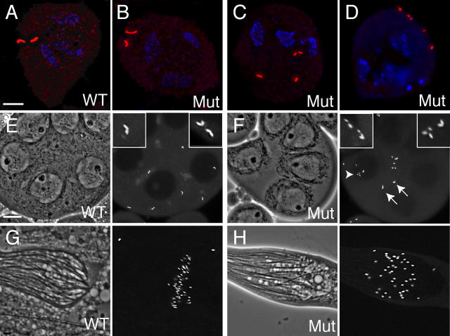
d-plp mutants exhibit centriole defects during spermatogenesis. (A–D) The distribution of centrioles (red; stained here with GTU88*) is shown in immature (A and B) or mature (C and D) WT (A) or mutant (C and D) primary spermatocytes. In immature spermatocytes, the centrioles in both WT (A) and mutant (B) cells are arranged as two orthogonal pairs, demonstrating that centriole replication has occurred normally during the mitotic divisions that generated these cells. This orthogonal arrangement is maintained in WT cells as they enter meiosis I (see Fig. 2 E), but in the majority of mutant cells (C and D) the centrioles lose their orthogonal arrangement and start to partially fragment. DNA is stained in blue. (E and F) Centriole fragmentation is also seen in living mutant spermatocytes. Phase-contrast images show part of a cyst of WT (E) or mutant (F) primary spermatocytes, whereas the fluorescent images show the distribution of centrioles (visualized here with GFP-PACT). Arrows highlight normal centriole pairs, arrowheads highlight centriole fragments. Insets show a 3×-magnified view of selected centrioles. (G) In WT cysts of maturing sperm, the centrioles/basal bodies (visualized with GFP-PACT) are located at the base of the sperm flagella. In mutant cysts of maturing sperm, basal bodies and flagella are present, but the cyst is often disorganized. Bars: 5 μm (A–D), 10 μm (E–H).

