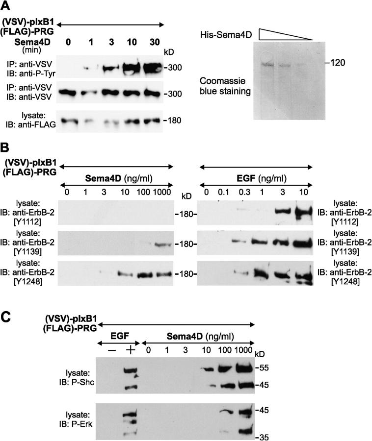
Figure 4.
Sema4D induces activation of ErbB-2. (A) HEK cells transfected with cDNAs encoding VSV-tagged plexin-B1 (VSV-plxB1) and FLAG-tagged PDZ-RhoGEF (FLAG-PRG) were treated with 100 ng/ml of purified Sema4D for the indicated time periods. VSV-tagged plexin-B1 was precipitated, and protein phosphorylation was visualized by antiphosphotyrosine antibodies. The right panel shows a Coomassie blue–stained gel with the preparation of purified Sema4D. (B) HEK293 cells were transfected with cDNAs encoding VSV-tagged plexin-B1 (VSV-plxB1) alone or together with FLAG-tagged PDZ-RhoGEF (FLAG-PRG). After 48 h, cells were starved, stimulated with EGF or Sema4D as indicated, and lysed. Phosphorylation site-specific antiphosphotyrosine antibodies directed against ErbB-2 tyrosine residues 1112, 1139, and 1248 were used to determine phosphorylation. (C) HEK293 cells were transfected as aforementioned, starved, stimulated with EGF or with increasing concentrations of Sema4D, and lysed. Specific antibodies directed against phosphorylated versions of Shc and Erk were used to visualize phosphorylation of those proteins.

