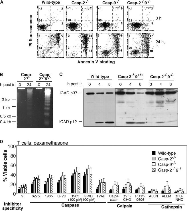
Figure 4.
Dying caspase-2 −/− 9 −/− cells display hallmarks of apoptosis and are protected by caspase inhibitors but not calpain/cathepsin inhibitors. (A–C) Ly5.1−CD4+8+ thymocytes sorted from reconstituted mice were cultured following 5 Gy γ irradiation (ir.) (A) Staining with FITC-conjugated Annexin V plus PI reveals phosphatidylserine exposure before loss of membrane integrity irrespective of genotype. (B) DNA “laddering” indicative of inter-nucleosomal fragmentation demonstrated by gel electrophoresis of genomic DNA. (C) Western blots showing processing of ICAD in dying caspase-2−/−9−/− cells. (D) The death of caspase-2−/−9−/− cells is antagonized by inhibitors of caspases but not of calpains or cathepsins. Ly5.1−Thy1+ mature T lymphocytes from the lymph nodes of reconstituted mice were cultured for 24 h in the presence of 100 nM dexamethasone plus the caspase inhibitors zVAD-fmk, IDN-6275 (6275), IDN-1965 (1965), or Q-VD-OPh (Q-VD), the calpain inhibitors z-VF-CHO or PD-150606, a cell permeable peptide of the calpain inhibitor calpastatin, the dual calpain/cathepsin inhibitors ALLM and ALLN or the cathepsin inhibitor z-FG-NHO-Bz-pOMe (zFG-NHO) (each at 50 μM). Cell death was quantified by PI staining and FACS analysis. Results represent mean ± SD of at least three independent experiments for each genotype. Vehicle: 0.4% DMSO.

