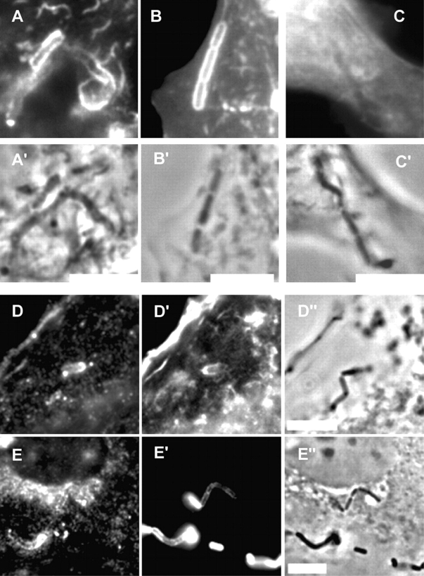
Figure 3.
Recruitment of raft markers during internalin-dependent entry. Non-transfected Lovo cells (A), Lovo cells transfected with CFP-MyrPalm (B), with CFP-GerGer (C), or with GFP-GPI (D), and epithelial Rov9 cells stably expressing the PrP (E), were incubated with L. innocua expressing internalin (BUG 1489) for 10 min, washed and fixed. The images B–D represent the CFP or GFP fluorescence, and the images A′, B′, C′, D′′, and E′′ correspond to phase contrast images. GM1 was labeled with fluorescent B subunit of cholera toxin (A). F-actin was labeled with Alexa Fluor 546 (D′). PrP (E) and internalin (E′) were labeled with mAbs (clone L7.7 and clone 4F2) and secondary fluorescent antibodies. Note that L. innocua expressing internalin, in some cases, appears as chains. Bars, 10 μm.

