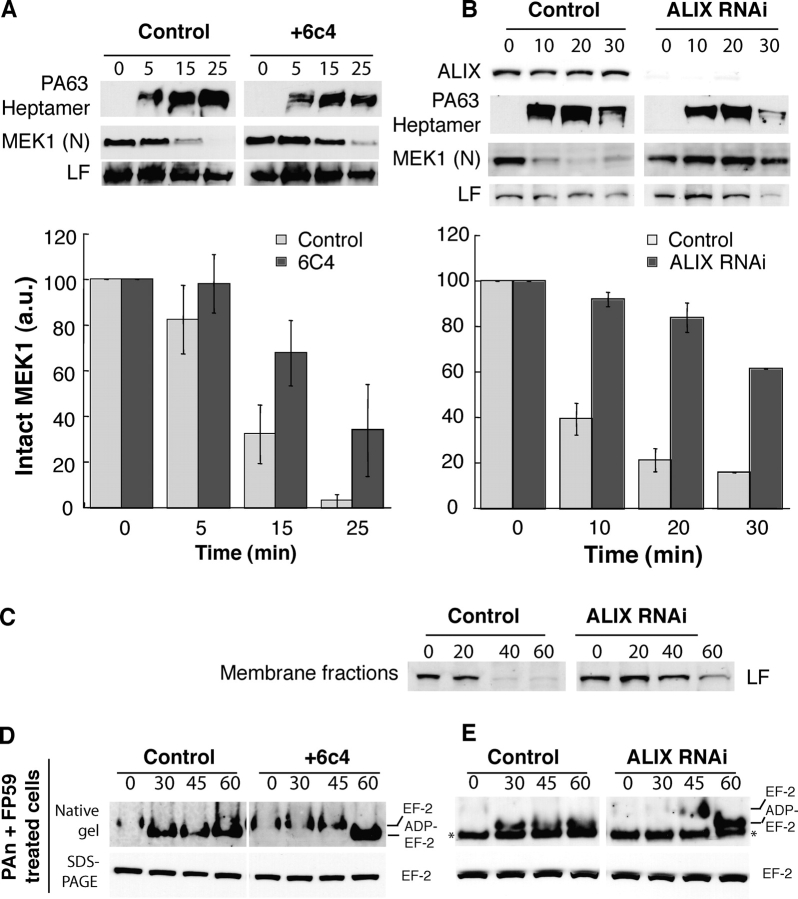
Figure 3.
Alteration of the dynamics of late endosomal intraluminal vesicles leads to a delay in MEK1 cleavage by LF. (A) CHO cells were incubated or not for 18 h with the anti-LBPA antibody 6c4 (50 μg/ml), washed, further incubated at 4°C for 1 h with 500 ng/ml PAn and 250 ng/ml LF, and transferred to 37°C for different periods of time (in min) in a toxin-free medium. 20 μg of PNS was analyzed by Western blotting to detect PAheptamer, the NH2 terminus of MEK1 and LF. The amount of intact MEK1 was quantified as in Fig. 2 A (n = 4). (B) HeLa cells were transfected with siRNA against ALIX, the efficiency of which was examined 78 h later by Western blotting using antibodies against ALIX. Cells were then incubated at 4°C for 1 h with 500 ng/ml PAn and 100 ng/ml LF, transferred to 37°C for different periods of time (in min) in a toxin-free medium, homogenized, and 20 μg of PNS was analyzed to detect the presence of PAheptamer and the NH2 terminus of MEK1. Quantifications were performed as in A. (C) HeLa cells were treated as in B. 80 μg of PNS were centrifuged at 100,000 g for 1 h and the pellet analyzed by Western blotting for LF. (D) CHO cells were treated as in A and incubated with 500 ng/ml PAn and 500 ng/ml FP59. 20 μg of cell lysates were loaded on native or SDS-PAGE before Western blotting against EF-2. (E) ALIX was knocked down in HeLa cells as in B, incubated with 500 ng/ml PAn with 500 ng/ml FP59, and the modification of EF2 analyzed as in D.

