
Figure 5.
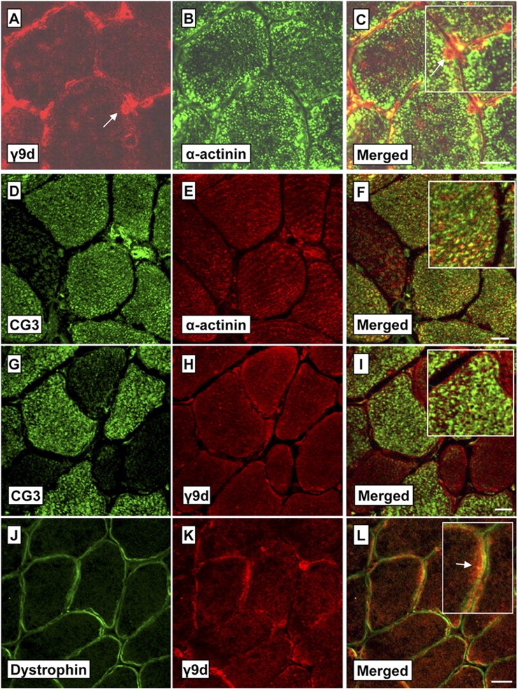
NS Tms recognized by the CG3 and γ9d antibodies have distinct myofiber localization. Shown are confocal images of transverse sections (7 μm) through soleus muscles stained with γ9d (A) or CG3 (D) and costained with α-actinin (B and E). The enlarged merged images (C and F, insets) indicate that the Tms recognized by γ9d and CG3 are not colocalized with α-actinin (labels myofibrils) and therefore are located outside the myofibrils. Further transverse sections (G–I) show that CG3 and γ9d stain separate regions within the myofiber (particularly notable in the enlarged inset, I). Cross sections costained with the membrane protein dystrophin (J) and γ9d (K) also show strong staining of γ9d at the myofiber periphery beneath the membrane (L, arrow in the enlarged inset). Bars, 20 μm.