Figure 4.
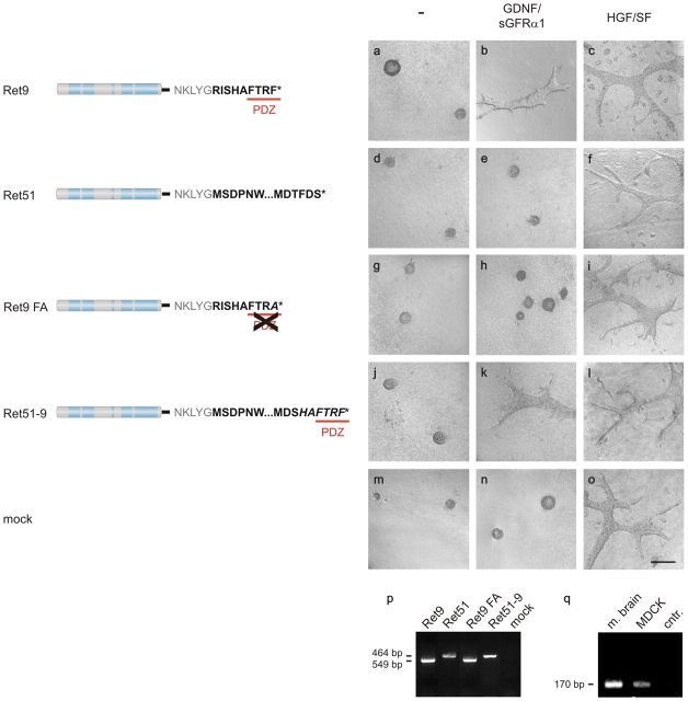
Induction of tubular structures by Ret depends on the PDZ-binding motif. MDCK cell lines expressing Ret9 (a–c), Ret51 (d–f), Ret9 FA (g–i), Ret51-9 (j–l), or control cells without Ret (m–o) were seeded as single cell suspensions into three-dimensional collagen matrices and stimulated with GDNF–sGFRα1 or HGF/SF. Photographs were taken after fixation. Bar in o: 200 μm (applies to a–o). (p) Expression of the Ret cDNA constructs in the various MDCK cell lines is shown by RT-PCR. (q) Expression of endogenous Shank3 in mouse brain and MDCK cells is shown by RT–PCR.

