Figure 8.
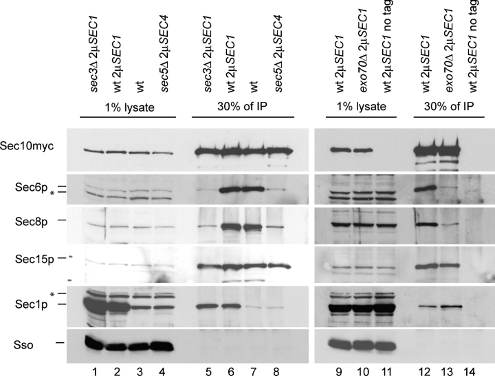
Co-isolation of Sec1p and exocyst subunits with myc epitope–tagged Sec10p. The exocyst was isolated from a wild-type (lanes 6, 7, and 12), sec3Δ (lane 5), sec5Δ (lane 8), and exo70Δ (lane 13) mutant strain via a carboxy-terminal 13myc epitope–tagged Sec10p. Immunoprecipitations were performed as described in Fig. 7. Mutant genotypes (wt, sec3Δ, sec5Δ, or exo70Δ) and the presence of multi-copy plasmids (2μSEC1 or 2μSEC4) are indicated on the top of each lane. 1% of lysates (left lanes) are compared with 30% of the immunoprecipitate (IP, right lanes). The antigens detected by Western blot analysis are marked on the left. Nonspecific bands are marked with an asterisk.

