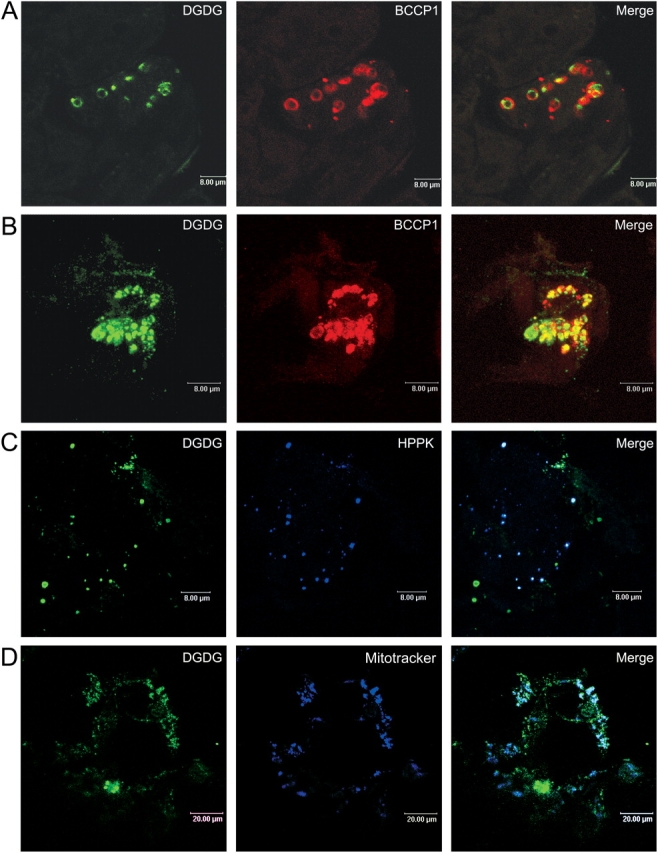
Figure 1.

Localization of DGDG in mitochondria of A. thalia na cells deprived of Pi for 3 d. Cells (A, control; B–D, Pi-deprived) were processed for indirect immunofluorescence labeling using anti-DGDG with secondary antibodies coupled to BODIPY and either anti-BCCP1, for chloroplast detection (A and B), or anti-HPPK (C), for mitochondria detection, with secondary antibodies coupled to Alexa 594. In D, mitochondria were visualized by staining with Mitotracker orange CMTMRos. Cells were observed by confocal microscopy. Bars: A–C, 8 μm; D, 20 μm.
