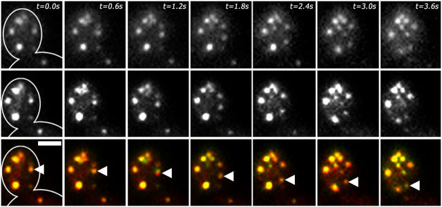
Figure 3.
Particles labeled with FM4-64 and Abp1p-GFP exhibit linear, retrograde movement. Mid-log phase wild-type haploid cells expressing Abp1p-GFP from the chromosomal locus were incubated with FM4-64 for 1 min at RT. Cells were washed with lactate medium to remove excess FM4-64, and imaged within 3 min after initial incubation with FM4-64. Two-color time-lapse imaging was performed as described for Fig. 1. Images shown are still frames from a time-lapse series showing Abp1p-GFP in the top row, FM4-64 in the middle row, and a merged image showing Abp1p-GFP in green and FM4-64 in red in the bottom row. The outline of the cell is shown in panels at t = 0 s. The bud, mother-bud neck, and part of the mother cell are shown. Arrowheads in the merged images mark an actin patch/endosome undergoing linear movement. Bar, 2 μm.

