Figure 2.
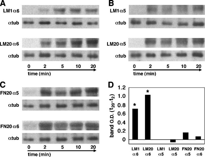
Cranial neural crest accumulate internalized surface integrin receptors in a substratum-dependent manner. Surface receptors were labeled with biotin, cells were incubated at 37°C for the indicated time points, then cooled to 25°C and treated with the reducing agent MesNa to remove biotin from receptors remaining on the surface. Cells were lysed, the receptors were immunoprecipitated, and the internalized portion revealed on Western blots with HRP-conjugated streptavidin. Bottom panels show internal loading control (α-tubulin). (A) On both low (LM1) and high (LM20) concentrations of laminin, there is an accumulation of internalized integrin α6 over 20 min. (B) On both LM1 and LM20, accumulation of internalized integrin α5 reaches steady-state levels by 2 min. (C) On high (FN20) fibronectin concentrations there is no accumulation of internalized integrin α5 or integrin α6 above the level observed at 2 min. (D) The average increase in band intensity from 2 to 20 min for each condition from three independent experiments is shown. Asterisks indicate significant increases in internalized α6 over time on both LM1 and LM20 (P < 0.05; Wilcoxon signed-rank test).

