
Figure 4.
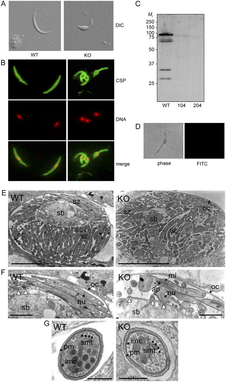
Characteristics of PbIMC1a-KO sporozoites. (A) Sporozoite shapes as observed by differential interference contrast (DIC). (B) Sporozoite shapes as observed by IFA staining for circumsporozoite protein (CSP, green) and DNA (red). (C) Western blot of oocyst sporozoites from WT and PbIMC1a-KO parasite clones 104 and 204. (D) Negative labeling of a PbIMC1a-KO sporozoite by IFA staining for PbIMC1a. (E) Cross section of sporulating oocysts. (F) Close-up view of E showing budding sporozoites. White arrowheads point at sporozoite budding sites. (G) Cross section of sporozoites. WT: wild-type, KO: PbIMC1a-KO, sb: sporoblast, sz: sporozoite, oc: oocyst wall, nu: nucleus, ap: apicoplast, mi: mitochondrion, pm: plasma membrane, imc: inner membrane complex, smt: subpellicular microtubules. Bars: 5 μm (E), 1 μm (F), 0.1 μm (G).