
Figure 6.
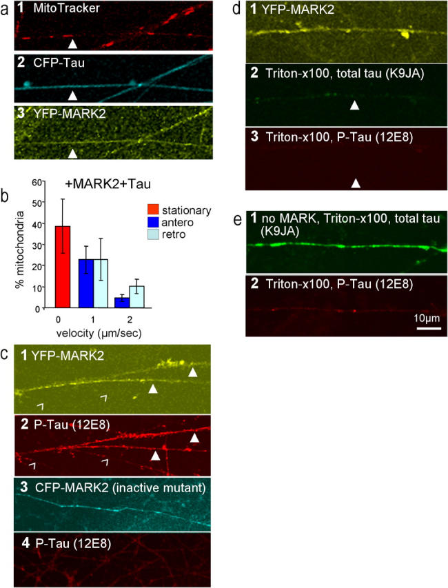
Recovery of anterograde movements of mitochondria by coexpression of tau and MARK2. (a,1–3) Axons of RGCs stained for mitochondria (MitoTracker, a 1), CFP-tau (a 2), YFP-MARK2 (a 3). Numerous mitochondria are present in axons, in spite of the presence of tau, because the phosphorylation of tau by MARK detaches it from microtubules and facilitates transport (closed arrowheads). (b) Velocity histogram of mitochondria. Note the substantial recovery of anterograde transport (dark blue bars) after coexpression with MARK2 (compare with Fig. 5 k, transfection of tau only). Error bars indicate SEM. (c, 1–4) Axons showing YFP-MARK2 (c 1) and phospho-Tau (at KXGS motifs, antibody 12E8, c 2, closed arrowheads). Axons after transfection with inactive CFP-MARK2 (c 3), which fails to increase the phosphorylation of KXGS motifs of tau (c 4, antibody 12E8 shows only background staining). (d, 1–3) Axons after transfection of cells with YFP-MARK2 (d 1) and then extracted with Triton X-100 and stained for total Tau (antibody K9JA, d 2) phospho-Tau (antibody 12E8, d 3). Note that Tau phosphorylated at KXGS motifs disappears because it is not attached to microtubules (closed arrowheads). (e, 1 and 2) Axons not transfected with MARK, extracted with Triton X-100, show strong staining with Tau (K9JA, e 1) but not with phospho-Tau (12E8, e 2).